About HepaRG™
HepaRG™ is a human bipotent progenitor cell line capable of differentiating into both biliary and hepatocyte lineages. It was originally derived from a female patient diagnosed with a cholangiocarcinoma and hepatitis C infection. The HepaRG cell line, free of hepatitis B and hepatitis C viruses has the following main characteristics:
- HepaRG cells can undergo a complete program of hepatocyte differentiation until a fully adult hepatic phenotype
- Hepatocyte-like cells are polarized cells able to form bile canaliculi-like structures
- Hepatocyte-like cells breath aerobically, consume lactate and contain as many mitochondria as the human hepatocytes
- HepaRG cell line has the potential to express major properties of stem cells: high plasticity & transdifferentiation capacity
The cells express the major nuclear receptors referred also as xeno-sensing receptors (Pregnane X receptor (PXR), constitutive androstane receptor (CAR), aryl hydrocarbon receptor (AHR)) (Figure 1, Antherieu et al., 2010 Aninat et al., 2005), drug and bile acids transporters (NTCP, BSEP, MRP2, MRP3, MRP4, MRP5, OSTa/b, MDR1, MDR3 etc.) (Figure 1 and 2, Bachour-El Azzi et al., 2015 and Anthérieuet al., 2010) and functional levels of phase I (CYP (CYP1A1/2, CYP2B6, CYP2Cs, CYP3A4 etc.) (Jinpeng Li et al. 2019) and II (UGT1A1, GSTA1, GSTA4, GSTM1) drug metabolizing enzymes (Figure 1, Anthérieu et al. 2010) as well as key hepatic nuclear factors such as Hepatocyte Nuclear Factor 4 (HNF4).
Furthermore, HepaRG™ cells show functional mitochondria, hepatokine secretion abilities and an adequate response to insulin (Figure 3). The HepaRG™ cell system appears as a robust surrogate for primary hepatocytes, which is versatile enough to study not only xenobiotic detoxification, but also the control of hepatic energy metabolism, secretory function and disease-related mechanisms (Tascher et al. 2019).
Unlike primary human hepatocytes, fully differentiated HepaRG cells can survive up to 4 weeks in culture allowing long-term studies and repeated exposures to drugs and chemicals (Figure 4, Jolly et al., 2020).
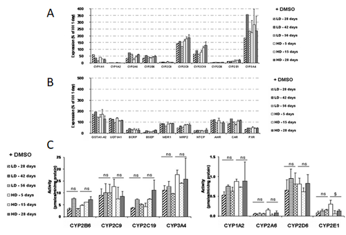
Figure 1. Basal mRNA levels of phase I and II metabolizing enzymes, membrane transporters, and nuclear receptors (A and B) and comparative basal activity levels of P450s (C) in HepaRG cells seeded at low density (LD) and cultured for 5 to 56 days or at high density (HD) and cultured for 5 to 28 days in presence of DMSO. Results are expressed as percentages compared with 1-day cultured human hepatocytes, arbitrarily set at 100% (Anthérieu et al., 2010).
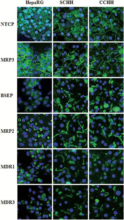
Figure 2. Distribution of main bile acid transporters in HepaRG cells and HHs.
Localization of several main hepatobiliary transporters was assessed by immunofluorescence staining in HepaRG cells as well as in 4–5 days HHs in sandwich (SCHH) and conventional (CCHH) cultures. Nuclei were labeled using Hoechst. Transporter immunolabeling in HepaRG cell cultures was restricted to hepatocyte-like cells; no transporter was visualized in primitive biliary cells (Bachour-El Azzi et al., 2015).
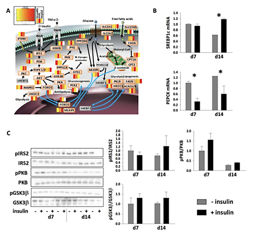
Figure 3. Insulin metabolism in highly differentiated HepaRG™ cells.
(A) The relative difference of protein abundances in HepaRG™ (defined serum-free medium: 0.5% DMSO, growth factor supplementation) and in HepG2 cells versus PHH, as shown as heatmaps for factors of insulin signaling/resistance pathways (white boxes: proteins not seen). Non-detected proteins are colored in grey. (B) mRNA levels of SREBP1c and PEPCK in differentiated HepaRG™ cells (differentiation medium: 1.7% DMSO, 10% FCS) in response to insulin stimulation (means ± SEM of three determinations). * indicates significant differences due to insulin stimulation for a given time point (ANOVA and post hoc Tukey tests; p < 0.05). (C) Phosphorylation status of IRS2, PKB, and GSK3β in differentiated HepaRG™ cells in response to insulin stimulation (means ± SEM of three determinations). (Tascher et al. 2019)
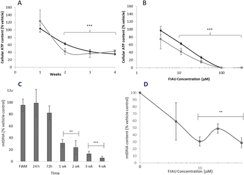
Figure 4. The utility of HepaRG cells in investigating the delayed toxicity.
Cellular ATP content after metabolic modification assay performed in HepaRG cells exposed to FIAU (0–300 μM). A: Time-response curve following exposure of HepaRG cells to 11 μM FIAU. B: Concentration-response curve following 2 weeks exposure of HepaRG cells. Round black markers represent data collected in glucose media, square black markers represent data collected in galactose media
Mitochondrial DNA content in HepaRG exposed to 10 μM FIAU (0–300 μM, 1–4 weeks). C: Time-response curve of FIAU following exposure to 10 μM FIAU or FIAM (4 weeks). B: Concentration-response curve following 2 weeks exposure to FIAU. Significance of each data point compared to vehicle control; ** p < .01, *** p < .001 (Jolly et al., 2020).
The process of HepaRG differentiation
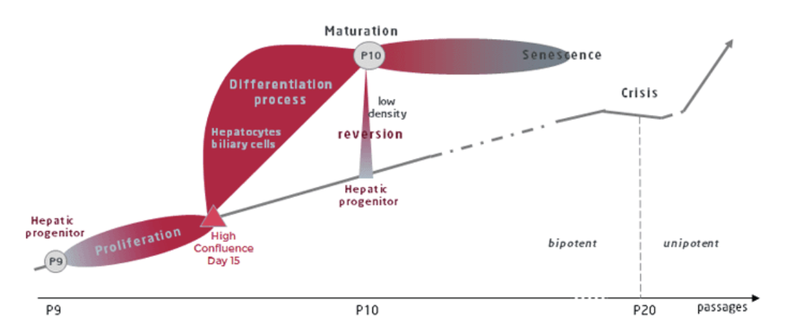
Two main stages are being identified, namely:
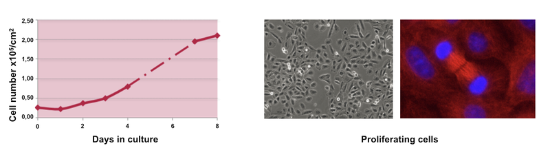
The proliferation stage
During the proliferation stage, the cell doubling time is 24h. At day 14, cells reach high confluence. At this moment the cells can be either split to re-enter proliferation stage or start the next differentiation stage.
The differentiation stage
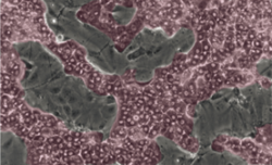
To undergo hepatic differentiation, the cells are shifted to a medium enriched supplemented with 1.7% DMSO for a 14-day period. Within 3-4 days, two distinctive cell populations are recognized: one corresponding to hepatocyte colonies and the other one corresponding to primitive biliary cells. Once fully differentiated, the hepatocyte-like cells express a full array of functions, responses, and regulatory pathways of primary human hepatocytes including: Phase I and II metabolizing-enzymes and transporter activities consistent with those found within a population of primary human hepatocytes including intact response elements, PXR, CAR and PPARa.
Confluent HepaRG™ cells start to commit into either hepatocyte (coloured in red) or biliary cells (coloured in grey).
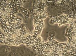
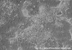
After 2 weeks, a mixed population of 2 types of cells, namely hepatocyte-like colonies surrounded by clear epithelial cells corresponding to primitive biliary cells, form a confluent monolayer which can be maintained in this stable form for several weeks in the presence of 1.7% DMSO.